

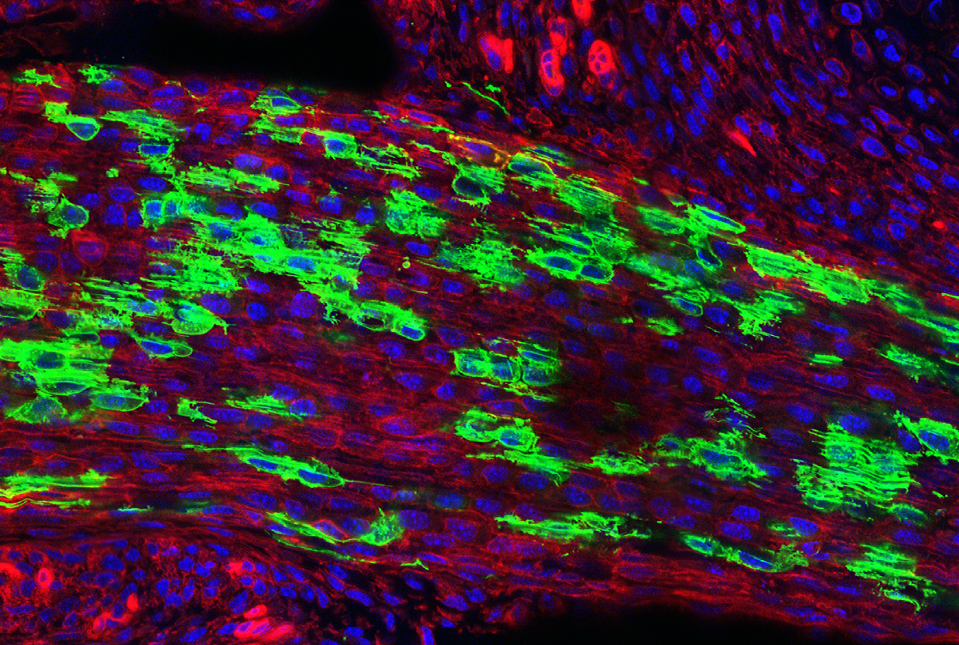
Researchers at Weill Cornell Medicine and Hospital for Special Surgery have discovered the stem cells—unspecialized cells that can develop into specific cell types—that give rise to the body’s tendons and ligaments, the tissues connecting muscles and bones. They also found that when these stem cells become hyperactive in the lower spine, they contribute to lumbar spinal stenosis. This condition, which impacts an estimated 103 million people worldwide, involves enlarged ligaments that can narrow the spinal canal and compress nerves, causing pain, numbness and difficulty walking.
The study, published Sept. 7 in Cell, suggests that therapeutics targeting these stem cells may lead to new treatment options for patients, including a class of drugs currently used to manage high blood pressure.
Dr. Matthew Greenblatt
“While previous studies had proposed several candidate stem cells, none had definitively shown that a single cell population could both self-renew and generate the full spectrum of tendon and ligament cell types,” said co-corresponding author Dr. Matthew Greenblatt, the Rohr Family Research Scholar, associate professor of pathology and laboratory medicine at Weill Cornell and a pathologist at NewYork-Presbyterian/Weill Cornell Medical Center.
Once the researchers had identified the elusive stem cells, they explored whether the findings could address an unmet clinical need for alternatives to surgery to treat lumbar spinal stenosis.
“Identifying these specialized stem cells unlocks a new area of research that allows us to address this disease much more mechanistically, rather than just waiting until a patient’s condition worsens and requires surgery to relieve the nerve compression,” said study co-corresponding author Dr. Sravisht Iyer, an associate professor of orthopedics at Weill Cornell and a spine surgeon at Hospital for Special Surgery (HSS). “The findings are exciting for their potential to change the way we deliver spinal care.”
Search for a Master Tendon and Ligament Stem Cell
Dr. Greenblatt was well equipped to lead the search. In 2018, he and his colleagues located the stem cell that initiates fracture repair in the outer layer of bone. They went on to identify the stem cells that form the skull and spine.

Dr. Sravisht Iyer
Unlike bone, however, tendons and ligaments are relatively uniform tissues with fibroblast-like cells that are difficult to distinguish from one another. As a result, finding a bona fide tendon and ligament stem-cell population was a challenge.
“We analyzed thousands of individual cells and sorted them into individual cell types. Then we identified which one had the properties we associate with ‘stemness,’” said Dr. Greenblatt. In other words, the rare cell population able to continually replenish itself and produce the mature cells needed to build and maintain these tissues. Studying mice, the researchers located the stem cells in a specialized anatomical niche within tendon and ligament tissue that serves as a reservoir for growth and repair.
Characterizing the mouse stem cell helped the researchers locate a human equivalent in ligament samples that Dr. Iyer had removed from patients during surgery with informed consent. The team—which included first author Dr. Lingling Hu, a postdoctoral fellow in Dr. Greenblatt’s and Dr. Iyer’s labs—confirmed that the human stem cells could both self-renew and produce ligament cells, demonstrating clinical relevance.
Furthermore, they determined that these stem cells are not relegated only to the spine. “We looked in the kneecap ligament; we looked at the Achilles tendon; and everywhere we looked, we found this cell,” said Dr. Greenblatt. “So, we think this is the universal stem cell for tendons and ligaments throughout the body.”
Potential Therapeutic Target for Spinal Stenosis
To find out if these cells are the culprits behind lumbar spinal stenosis, the researchers compared stem cells harvested from individuals with lumbar spinal stenosis to those isolated from spinal ligaments removed from people with herniated discs and no signs of stenosis. The patients provided informed consent pre-operatively.
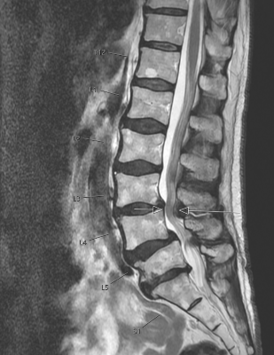
MRI image of spinal stenosis with arrow indicating narrowing of the spinal canal. Credit: Dr. Sravisht Iyer
The researchers detected higher stem cell numbers in the ligaments taken from people with stenosis. When these stenosis-derived cells were transplanted into mice, they produced more tendon cells than healthy stem cells did. “Though spinal stenosis is a complex condition, this really showed us that these cells are contributing to the pathology,” said Dr. Greenblatt, who is also a member of the Sandra and Edward Meyer Cancer Center at Weill Cornell.
On a molecular level, the stenosis-associated stem cells showed higher levels of calcium signaling than their healthy counterparts. In fact, the researchers could trigger tissue overgrowth by increasing calcium signaling in stem cells from healthy ligaments using genetic manipulation.
Just as important, the team showed that curtailing calcium signaling blocked cell overgrowth in a mouse model of lumbar spinal stenosis. This may indicate that calcium channel blockers currently used to treat high blood pressure could be repurposed to treat spinal stenosis, but clinical studies will be needed to explore this direction.
“This is probably the first work that's shown a potential therapeutic target for one of the most common spine conditions in the world,” said Dr. Iyer.
Dr. Lingling Hu
Beyond lumbar spinal stenosis, Dr. Greenblatt hopes to explore the role that these stem cells might play in other conditions, such as Marfan syndrome, a genetic disorder that affects the body’s connective tissues.
These findings could also eventually help researchers find treatments for tendons and ligaments that heal poorly after injuries, including rotator cuff tears, Achilles tendon injuries, ligament reconstruction and chronic tendon degeneration.
“Given that this cell appears to be the ultimate origin of all tendon and ligament cells, defects in this cell are likely at the heart of a wide range of tendon and ligament disorders,” said Dr. Greenblatt.
This work was supported in part by the Marfan Foundation’s Victor A. McKusick Fellowship, a Kellen Scholars Award, the Children’s Tumor Foundation (CTF-2023-01-005), the Arthritis National Research Foundation (1065843), a Jumpstart award from Weill Cornell, a National Research Foundation of Korea (NRF) award funded by the Ministry of Education (NRF-2021R1A6A3A14038667), the National Institutes of Health (grants T32-AR078751 and T32-AR071302-07), a MIND Prize from the Pershing Square Foundation, a Mary Kay Ash Foundation Award, an Innovator Award from the Marfan Foundation and a Burroughs Wellcome Career Award for Medical Scientists.

